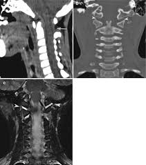
The anatomy of the craniovertebral junction, although complex, may be well visualized by routine MR imaging. This essay discusses the anatomy of the complex articulations of the craniovertebral junction. Representative MR images and gross anatomic photographs are presented to illustrate the intricate ligamentous and articular anatomy. Knowledge of the normal anatomy of the occipitoatlantoaxial region is necessary in order to understand the common disorders that affect this area. The most common disorders are trauma and arthropathies, but also include congenital abnormalities and neoplasm. The resultant abnormal mechanics may lead to neurologic sequelae or pain


 Contact Us


 Hospitals
 Doctors
 Diagnostic
 Pharmacy
 Health Tips
 Blog
Blog


Comments