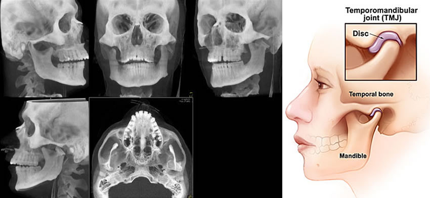
The purpose of this study was to correlate disc position and the type of disc displacement, intra-capsular effusion and degenerative changes of the condyle as demonstrated in MRI studies. In this study, 126 temporomandibular joints (TMJs) of 63 patients with TMJ disorders were investigated using clinical examination and MRI. One hundred and twelve TMJs were found to have internal derangement as disc displacement. The angle between the posterior margin of the disc and the vertical line drawn through the centre of the condyle was measured on MRI for each TMJ. The positions of the discs were normal, 0 degrees-10 degrees, in 11.11%; slightly displaced, 11 degrees-30 degrees, in 37.30%; mildly displaced 31 degrees-50 degrees, in 15.08%; moderately displaced, 51 degrees-80 degrees, in 7.14% of the TMJs with anterior displacement with reduction (ADDR). The disc position was severely displaced anteriorly, as over 80 degrees, in all TMJs with anterior disc displacement without reduction (ADD), constituting 27.78% of all cases. We found that the smaller the degree of disc displacement the milder the internal derangement and that the intra-capsular effusion was more frequently associated with TMJ with ADDR. The degenerative condylar changes were more severe with an increased degree of anterior disc displacement.


 Contact Us


 Hospitals
 Doctors
 Diagnostic
 Pharmacy
 Health Tips
 Blog
Blog


Comments