Find out health conditions and services

Find out health conditions and services

KayaWell Icon
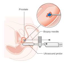
Ulcerative colitis (UC) is a long-term condition that results in inflammation and ulcers of the colon and rectum. The primary symptom of active disease is abdominal pain and diarrhea mixed with blood. Weight loss, fever, and anemia may also occur. Often symptoms come on slowly and can range from mild to severe. Symptoms typically occur intermittently with periods of no symptoms between flares. Complications may include megacolon, inflammation of the eye, joints, or liver, and colon cancer.

The cause of UC is unknown. Theories involve immune system dysfunction, genetics, changes in the normal gut bacteria, and environmental factors. Rates tend to be higher in the developed world with some proposing this to be the result of less exposure to intestinal infections, or a Western diet and lifestyle. The removal of the appendix at an early age may be protective. Diagnosis is typically by colonoscopy with tissue biopsies. It is a kind of inflammatory bowel disease (IBD) along with Crohn's disease and microscopic colitis.
Treatment Idea Insurance Policy Near By Expert Wellness Plan Events Community Forum Wiki
Indigestion, also known as dyspepsia, is a condition of impaired digestion. Symptoms may include upper abdominal fullness, heartburn, nausea, belching, or upper abdominal pain. People may also experience feeling full earlier than expected when eating. Dyspepsia is a common problem and is frequently caused by gastroesophageal reflux disease (GERD) or gastritis. In a small minority of cases it may be the first symptom of peptic ulcer disease (an ulcer of the stomach or duodenum) and, occasionally, cancer. Hence, unexplained newly onset dyspepsia in people over 55 or the presence of other alarming symptoms may require further investigations.

Functional indigestion (previously called nonulcer dyspepsia) is indigestion "without evidence of an organic disease that is likely to explain the symptoms". Functional indigestion is estimated to affect about 15% of the general population in western countries.
Treatment Idea Insurance Policy Near By Expert Wellness Plan Events Community Forum Wiki
Uric acid is a heterocyclic compound of carbon, nitrogen, oxygen, and hydrogen with the formula C5H4N4O3. It forms ions and salts known as urates and acid urates, such as ammonium acid urate. Uric acid is a product of the metabolic breakdown of purine nucleotides, and it is a normal component of urine. High blood concentrations of uric acid can lead to gout and are associated with other medical conditions including diabetes and the formation of ammonium acid urate kidney stones.
Treatment Idea Insurance Policy Near By Expert Wellness Plan Events Community Forum Wiki
Urinary incontinence (UI), also known as involuntary urination, is any leakage of urine. It is a common and distressing problem, which may have a large impact on quality of life. It is twice as common in women as in men. Pregnancy, childbirth, and menopause are major risk factors. It has been identified as an important issue in geriatric health care. Urinary incontinence is often a result of an underlying medical condition but is under-reported to medical practitioners. Enuresis is often used to refer to urinary incontinence primarily in children, such as nocturnal enuresis (bed wetting).

There are four main types of incontinence:

Urge incontinence due to an overactive bladder
Stress incontinence due to poor closure of the bladder
Overflow incontinence due to either poor bladder contraction or blockage of the urethra
Functional incontinence due to medications or health problems making it difficult to reach the bathroom
Treatment Idea Insurance Policy Near By Expert Wellness Plan Events Community Forum Wiki
A urinary tract infection (UTI) is an infection that affects part of the urinary tract. When it affects the lower urinary tract it is known as a bladder infection (cystitis) and when it affects the upper urinary tract it is known as kidney infection (pyelonephritis). Symptoms from a lower urinary tract include pain with urination, frequent urination, and feeling the need to urinate despite having an empty bladder. Symptoms of a kidney infection include fever and flank pain usually in addition to the symptoms of a lower UTI. Rarely the urine may appear bloody. In the very old and the very young, symptoms may be vague or non-specific.

The most common cause of infection is Escherichia coli, though other bacteria or fungi may rarely be the cause. Risk factors include female anatomy, sexual intercourse, diabetes, obesity, and family history. Although sexual intercourse is a risk factor, UTIs are not classified as sexually transmitted infections (STIs). Kidney infection, if it occurs, usually follows a bladder infection but may also result from a blood-borne infection. Diagnosis in young healthy women can be based on symptoms alone. In those with vague symptoms, diagnosis can be difficult because bacteria may be present without there being an infection. In complicated cases or if treatment fails, a urine culture may be useful.
Treatment Idea Insurance Policy Near By Expert Wellness Plan Events Community Forum Wiki

A urobilinogen in urine test measures the amount of urobilinogen in a urine sample. Urobilinogen is formed from the reduction of bilirubin. Bilirubin is a yellowish substance found in your liver that helps break down red blood cells. Normal urine contains some urobilinogen. If there is little or no urobilinogen in urine, it can mean your liver isn't working correctly. Too much urobilinogen in urine can indicate a liver disease such as hepatitis or cirrhosis.

Other names: urine test; urine analysis; UA, chemical urinalysis

Treatment Idea Insurance Policy Near By Expert Wellness Plan Events Community Forum Wiki

Uterine fibroids are benign tumors that originate in the uterus (womb). Although they are composed of the same smooth muscle fibers as the uterine wall (myometrium), they are much denser than normal myometrium. Uterine fibroids are usually round.Uterine fibroids are often described based upon their location within the uterus. Subserosal fibroids are located beneath the serosa (the lining membrane on the outside of the uterus). These often appear localized on the outside surface of the uterus or may be attached to the outside surface by a pedicle. Submucosal (submucous) fibroids are located inside the uterine cavity beneath the inner lining of the uterus. Intramural fibroids are located within the muscular wall of the uterus.

Treatment Idea Insurance Policy Near By Expert Wellness Plan Events Community Forum Wiki

Uterine polyps are soft red outgrowths from the lining of the womb (the endometrium), usually less than 1 cm in diameter, which often flatten to fit the cavity of the uterus.The stalk of the polyp (or pedicle) is usually short, but sometimes it grows long enough for the polyp to project from the cervix (the lower opening of the womb). Polyps are prone to bleeding, and a uterine polyp that develops near the fallopian tubes may obstruct the opening of the tubes, possibly leading to difficulty with becoming pregnant. Uterine polyps can develop in pre- or post-menopausal women. Very rarely, polyps can be cancerous.


Treatment Idea Insurance Policy Near By Expert Wellness Plan Events Community Forum Wiki

The uterus (the womb, in which a fetus develops) is normally held in place inside the pelvis with various muscles and ligaments. Sometimes, because of childbirth or difficult labor and vaginal delivery, these tissues are weakened. As a woman ages and with age-related decrease in the concentration of the hormone estrogen, her uterus can move downward into the vaginal canal, causing the condition known as a prolapsed uterus.Muscle weakness or relaxation may allow the uterus to sag or come completely out of the body. Prolapsed uterus can be described in the following stages

Treatment Idea Insurance Policy Near By Expert Wellness Plan Events Community Forum Wiki

Although it is a relatively rare disease, primarily found in the Caucasian population, uveal melanoma is the most common primary intraocular tumor in adults with a mean age-adjusted incidence of 5.1 cases per million per year. Tumors are located either in iris (4%), ciliary body (6%), or choroid (90%). The host susceptibility factors for uveal melanoma include fair skin, light eye color, inability to tan, ocular or oculodermal melanocytosis, cutaneous or iris or choroidal nevus, and BRCA1-associated protein 1 mutation. Currently, the most widely used first-line treatment options for this malignancy are resection, radiation therapy, and enucleation. There are two main types of radiation therapy: plaque brachytherapy (iodine-125, ruthenium-106, or palladium-103, or cobalt-60) and teletherapy (proton beam, helium ion, or stereotactic radiosurgery using cyber knife, gamma knife, or linear accelerator). The alternative to radiation is enucleation. Although these therapies achieve satisfactory local disease control, long-term survival rate for patients with uveal melanoma remains guarded, with risk for liver metastasis. There have been advances in early diagnosis over the past few years, and with the hope survival rates could improve as smaller tumors are treated. As in many other cancer indications, both early detection and early treatment could be critical for a positive long-term survival outcome in uveal melanoma. These observations call attention to an unmet medical need for the early treatment of small melanocytic lesions or small melanomas in the eye to achieve local disease control and vision preservation with the possibility to prevent metastases and improve overall patient survival.

Treatment Idea Insurance Policy Near By Expert Wellness Plan Events Community Forum Wiki

A urinary tract infection (UTI) is an infection in any part of your urinary system — your kidneys, ureters, bladder and urethra. Most infections involve the lower urinary tract — the bladder and the urethra.Women are at greater risk of developing a UTI than are men. Infection limited to your bladder can be painful and annoying. However, serious consequences can occur if a UTI spreads to your kidneys.Doctors typically treat urinary tract infections with antibiotics. But you can take steps to reduce your chances of getting a UTI in the first place

Treatment Idea Insurance Policy Near By Expert Wellness Plan Events Community Forum Wiki

An umbilical hernia creates a soft swelling or bulge near the navel (umbilicus). If your baby has an umbilical hernia, you may notice the bulge only when he or she cries, coughs or strains. The bulge may disappear when your baby is calm or lies on his or her back.Umbilical hernias in children are usually painless. Umbilical hernias that appear during adulthood may cause abdominal discomfort.

Treatment Idea Insurance Policy Near By Expert Wellness Plan Events Community Forum Wiki

Puffiness under the eyes can be bothersome, but it’s usually just a cosmetic concern. While rare, undereye puffiness can be a symptom of some kidney and thyroid conditions. If you occasionally wake up with puffiness under the eyes, consider what you did the day before to look for lifestyle causes such as staying up late or eating too much salt. Consider talking to your doctor about possible treatments for chronic undereye puffiness.

Treatment Idea Insurance Policy Near By Expert Wellness Plan Events Community Forum Wiki

Unstable angina belongs to the spectrum of clinical presentations referred to collectively as acute coronary syndromes (ACSs), which also includes ST-segment elevation myocardial infarction (STEMI) and non-STEMI (NSTEMI). [1, 2] Unstable angina is considered to be an ACS in which there is myocardial ischemia without detectable myocardial necrosis (ie, cardiac biomarkers of myocardial necrosis —such as creatine kinase MB isozyme, troponin, myoglobin—are not released into the circulation). See the image below.

Treatment Idea Insurance Policy Near By Expert Wellness Plan Events Community Forum Wiki

Cancer of the ureter (ureteral cancer) is a type of cancer that begins in the cells that line the inside of the tubes (ureters) that connect your kidneys to your bladder. Ureters are part of the urinary tract, and they carry urine produced by the kidneys to the bladder.Ureteral cancer is uncommon. It occurs most often in older adults and in people who have previously been treated for bladder cancer.Ureteral cancer is closely related to bladder cancer. The cells that line the ureters are the same type of cells that line the inside of the bladder. People diagnosed with ureteral cancer have a greatly increased risk of bladder cancer, so your doctor will recommend tests to look for signs of bladder cancer.Treatment for ureteral cancer typically involves surgery. In certain cases, chemotherapy may be recommended.

Treatment Idea Insurance Policy Near By Expert Wellness Plan Events Community Forum Wiki

Ureteral obstructions are blockages in your urinary tract, which includes your kidneys, bladder, the tubes that carry urine from your kidneys to your bladder (ureters), and the tube that connects your bladder to the outside of your body (urethra). Blockages can develop for many reasons, including gastrointestinal problems. Ureteral obstructions are more common in men, especially as they get older and their prostate gland enlarges.Obstructions can be cured with surgery, but they need to be treated promptly. If they’re not, they can lead to severe illness, kidney damage and life-threatening infections.

Treatment Idea Insurance Policy Near By Expert Wellness Plan Events Community Forum Wiki

Ultrasound imaging also called as sonography uses a transducer or probe to generate sound waves and produce pictures of the body's internal structures. It is often used to help diagnose unexplained pain, swelling or infection. It is used to see internal body structures such as tendons, muscles, joints, vessels and internal organs. It may also be used to provide imaging guidance to needle biopsies or to see and evaluate conditions related to blood flow. It is also the preferred imaging method for monitoring a pregnant woman and her unborn child. It does not use ionizing radiation, has no known harmful effects, and provides a clear picture of soft tissues that don't show up well on x-ray images.Most of these level II ultrasounds should be done in the second trimester of pregnancy usually between 18 and 22 weeks. It uses sound waves to produce pictures of a baby (embryo or fetus) within a pregnant woman, as well as the mother's uterus and ovaries. It does not use ionizing radiation, has no known harmful effects, and is the preferred method for monitoring pregnant women and their unborn babies. A Doppler ultrasound study – a technique that evaluates blood flow in the umbilical cord, fetus or placenta – may be part of this exam. It is a totally painless and safe procedure. It is useful test to establish the presence of fetus, estimate the age of pregnancy, diagnose congenital abnormalities of the fetus, assess fetal growth and well being etc. Preparation - No special preparation is needed.


Treatment Idea Insurance Policy Near By Expert Wellness Plan Events Community Forum Wiki

Your doctor has requested an ultrasound-guided biopsy. The goal of a biopsy is to remove a sample of tissue for testing in a laboratory. A biopsy can help diagnose abnormalities such as infection, inflammation or malignancy. During your biopsy, an imaging physician will use an ultrasound scanner to accurately guide a needle to the site of the biopsy. The needle will then be used to remove a tissue sample. At the S. Mark Taper Foundation Imaging Center, a highly trained medical team will oversee your procedure. This team will include an imaging physician, a diagnostic medical sonographer and a registered nurse.


Treatment Idea Insurance Policy Near By Expert Wellness Plan Events Community Forum Wiki

An ultrasound / SONOGRAPHY is a procedure that uses high-frequency sound waves to scan a the internal organs of the body woman’s abdomen and pelvic cavity, the reproductive system and the fetus of a pregnant woman creating a picture (sonogram) of the baby and placenta. Although the terms ultrasound and sonogram are technically different, they are used interchangeably and reference the same exam. They can show the structure and movement of the body's internal organs, including the abdomen, the fetus of pregnant women as well as blood flowing through blood vessels.

Treatment Idea Insurance Policy Near By Expert Wellness Plan Events Community Forum Wiki

A biophysical profile (BPP) test measures the health of your baby (fetus) during pregnancy. A BPP test may include a non stress test with electronic fetal heart monitoring and a fetal ultrasound. The BPP measures your baby's heart rate, muscle tone, movement, breathing, and the amount of amniotic fluid around your baby. A BPP is commonly done in the last trimester of pregnancy. If there is a chance that your baby may have problems during your pregnancy (high-risk pregnancy), a BPP may be done by 32 to 34 weeks or earlier. Some women with high-risk pregnancies may have a BPP test every week or twice a week in the third trimester. It is usually done to keep track of your baby’s health. 

Treatment Idea Insurance Policy Near By Expert Wellness Plan Events Community Forum Wiki

A breast ultrasound uses sound waves to make a picture of the tissues inside the breast. A breast ultrasound can show all areas of the breast, including the area closest to the chest wall, which is hard to study with a mammogram. Breast ultrasound does not use X-rays or other potentially harmful types of radiation. A breast ultrasound is used to see whether a breast lump is filled with fluid (a cyst) or if it is a solid lump. An ultrasound does not replace the need for a mammogram, but it is often used to check abnormal results from a mammogram


Treatment Idea Insurance Policy Near By Expert Wellness Plan Events Community Forum Wiki

Ultrasound imaging of the breast uses sound waves to produce pictures of the internal structures of the breast. It’s primarily used to help diagnose breast lumps or other abnormalities your doctor may have found during a physical exam, mammogram or breast MRI. Ultrasound is safe, noninvasive and does not use ionizing radiation.This procedure requires little to no special preparation. Leave jewelry at home and wear loose, comfortable clothing. You will be asked to undress from the waist up and to wear a gown during the procedure.

Treatment Idea Insurance Policy Near By Expert Wellness Plan Events Community Forum Wiki

 Ultrasound imaging of the head uses sound waves to produce pictures of the brain and cerebrospinal fluid. It is most commonly performed on infants, whose skulls have not completely formed. A transcranial Doppler ultrasound evaluates blood flow in the brain’s major arteries. Ultrasound is safe, noninvasive, and does not use ionizing radiation.This procedure requires little to no special preparation. Your doctor will instruct you on how to prepare, including whether adults undergoing the exam should refrain from using nicotine-based products that may cause blood vessels to constrict. Leave jewelry at home and wear loose, comfortable clothing. You may be asked to wear a gown.

Treatment Idea Insurance Policy Near By Expert Wellness Plan Events Community Forum Wiki

 B-scan ultrasonography is an important adjuvant for the clinical assessment of various ocular and orbital diseases. With understanding of the indications for ultrasonography and proper examination technique, one can gather a vast amount of information not possible with clinical examination alone. This article is designed to describe the principles, techniques, and indications for echographic examination, as well as to provide a general understanding of echographic characteristics of various ocular pathologies. 

Treatment Idea Insurance Policy Near By Expert Wellness Plan Events Community Forum Wiki

USG scan for Follicular Monitoring is done to study ovarian follicles that are used to identify ovulation of egg. For couples planning for pregnancy, ovulation period is the important period to try on, for conception to happen. Out of many ways available, the most reliable way to understand the ovulation is Follicular monitoring. It’s an endovaginal scan carried to study the ovaries, uterus and uterus lining. Looking at ovaries, the growth of follicles inside ovaries can be assessed giving a chance to predict the ovulation and also look if the womb is getting ready for pregnancy.

With follicular monitoring, you will get to know the growth of follicles, the approximate rupture period of follicles and hence can predict the ovulation period thereby have an intercourse happen at the right time for conception to take place. The womb if not yet ready for pregnancy can be treated by some supplements prescribed by the doctors.

This scan is carried out in a slightly dark examination room. You will have to undress down from your waist while you get covered by a sheet. Now you will be asked to position yourself lying down on your back on a special bed with your knees bent. Once you position yourself comfortably; the transducer, a sterile lubricated covered electronic device, is gently inserted into your vagina to look at the uterus and ovaries. Depending on how relaxed the patient is, the test takes up to 15 minutes.


Treatment Idea Insurance Policy Near By Expert Wellness Plan Events Community Forum Wiki

 X-ray beams are passed through the abdomen, producing images of the kidneys, ureters, and bladder on a special type of film. KUB radiography is often used as a first step in diagnosing problems of the urinary system, and is usually done in conjunction with intravenous pyelography.


Treatment Idea Insurance Policy Near By Expert Wellness Plan Events Community Forum Wiki

An ultrasound / SONOGRAPHY is a procedure that uses high-frequency sound waves to scan a the internal organs of the body woman’s abdomen and pelvic cavity, the reproductive system and the fetus of a pregnant woman creating a picture (sonogram) of the baby and placenta. Although the terms ultrasound and sonogram are technically different, they are used interchangeably and reference the same exam. They can show the structure and movement of the body's internal organs, including the abdomen, the fetus of pregnant women as well as blood flowing through blood vessels.

Treatment Idea Insurance Policy Near By Expert Wellness Plan Events Community Forum Wiki

Thyroid ultrasound uses sound waves to produce pictures of the thyroid gland within the neck. It does not use ionizing radiation and is commonly used to evaluate lumps or nodules found during a routine physical or other imaging exam.This procedure requires little to no special preparation. Leave jewelry at home and wear loose, comfortable clothing. You may be asked to wear a gown.

Treatment Idea Insurance Policy Near By Expert Wellness Plan Events Community Forum Wiki

Ultrasound  is safe and paUltrasoundinless, and produces pictures of the inside of the body using sound waves. Ultrasound imaging, also called ultrasound scanning or sonography, involves the use of a small transducer (probe) and ultrasound gel placed directly on the skin. High-frequency sound waves are transmitted from the probe through the gel into the body. The transducer collects the sounds that bounce back and a computer then uses those sound waves to create an image. Ultrasound examinations do not use ionizing radiation (as used in x-rays), thus there is no radiation exposure to the patient. Because ultrasound images are captured in real-time, they can show the structure and movement of the body's internal organs, as well as blood flowing through blood vessels.


Treatment Idea Insurance Policy Near By Expert Wellness Plan Events Community Forum Wiki

Ultrasound is a high-frequency sound that you cannot hear but it travels through fluid and soft tissues. However, it bounces back, or echoes, when it hits a more solid, dense surface such as a valve or bile in the gallbladder etc. So, when ultrasound 'hits' structures of different densities in our bodies, it echoes differently with each hit. 


Treatment Idea Insurance Policy Near By Expert Wellness Plan Events Community Forum Wiki

Ultrasound imaging of the scrotum uses sound waves to produce pictures of a man’s testicles and surrounding tissues. It is the primary method used to help evaluate disorders of the testicles, epididymis (a tube immediately next to a testicle that collects sperm) and scrotum. Ultrasound is safe, noninvasive, and does not use ionizing radiation.

Treatment Idea Insurance Policy Near By Expert Wellness Plan Events Community Forum Wiki

An ultrasound / SONOGRAPHY is a procedure that uses high-frequency sound waves to scan a the internal organs of the body woman’s abdomen and pelvic cavity, the reproductive system and the fetus of a pregnant woman creating a picture (sonogram) of the baby and placenta. Although the terms ultrasound and sonogram are technically different, they are used interchangeably and reference the same exam. They can show the structure and movement of the body's internal organs, including the abdomen, the fetus of pregnant women as well as blood flowing through blood vessels


Treatment Idea Insurance Policy Near By Expert Wellness Plan Events Community Forum Wiki

Thyroid ultrasound uses sound waves to produce pictures of the thyroid gland within the neck. It does not use ionizing radiation and is commonly used to evaluate lumps or nodules found during a routine physical or other imaging exam.This procedure requires little to no special preparation. Leave jewelry at home and wear loose, comfortable clothing. You may be asked to wear a gown.

Treatment Idea Insurance Policy Near By Expert Wellness Plan Events Community Forum Wiki

A transrectal ultrasound (TRUS) may also be called prostate sonogram or endorectal ultrasound. It is used to look at the prostate and tissues around it. An ultrasound transducer (also called a probe) sends sound waves through the wall of the rectum and into the prostate and surrounding tissue. A computer analyzes the wave patterns (called echoes) as they bounce off the organs and converts them into an image that doctors view on a video screen.

Treatment Idea Insurance Policy Near By Expert Wellness Plan Events Community Forum Wiki

An ultrasound test uses high-frequency sound waves to create images of your internal organs. Imaging tests can identify abnormalities and help doctors diagnose conditions. A transvaginal ultrasound, also called an endovaginal ultrasound, is a type of pelvic ultrasound used by doctors to examine female reproductive organs. This includes the uterus, fallopian tubes, ovaries, cervix, and vagina.

“Transvaginal” means “through the vagina.” This is an internal examination. Unlike a regular abdominal or pelvic ultrasound, where the ultrasound wand, or transducer, rests on the outside of the pelvis, this procedure involves your doctor or a technician inserting an ultrasound probe about two or three inches into your vaginal canal.


Treatment Idea Insurance Policy Near By Expert Wellness Plan Events Community Forum Wiki

An ultrasound / SONOGRAPHY is a procedure that uses high-frequency sound waves to scan a the internal organs of the body woman’s abdomen and pelvic cavity, the reproductive system and the fetus of a pregnant woman creating a picture (sonogram) of the baby and placenta. Although the terms ultrasound and sonogram are technically different, they are used interchangeably and reference the same exam. They can show the structure and movement of the body's internal organs, including the abdomen, the fetus of pregnant women as well as blood flowing through blood vessels.


Treatment Idea Insurance Policy Near By Expert Wellness Plan Events Community Forum Wiki
Ultrasound imaging also called as sonography uses a transducer or a probe to generate sound waves and produce pictures of the body's internal structures. It is often used to help diagnose unexplained pain, swelling or infection. It is also used to see internal body structures such as tendons, muscles, joints, vessels and internal organs. Ultrasound abdomen is primarily used for evaluating the kidneys, liver, pancreas, gall bladder etc.It may also be used to provide imaging guidance to needle biopsies or to see and evaluate conditions related to blood flow. It does not use ionizing radiation, has no known harmful effects, and provides a clear picture of soft tissues that don't show up well on x-ray images. It is a painless, safe and non invasive procedure.
Treatment Idea Insurance Policy Near By Expert Wellness Plan Events Community Forum Wiki